Revision Rhinoplasty: What Can Be Corrected If You Are Unhappy With the Initial Result?
 All articles
Rhinoplasty does not always deliver the result a patient hoped for. In some cases, aesthetic concerns remain after surgery, the shape of the nasal tip changes, or breathing difficulties develop. Revision rhinoplasty may be considered in such situations. However, it requires particularly careful planning, as the surgeon must work with tissues that have already undergone structural changes.
What Is Revision Rhinoplasty?
Revision rhinoplasty, also known as secondary rhinoplasty, is a procedure performed to correct the shape of the nose and restore its function following a previous rhinoplasty. It may be recommended when the initial result does not meet the patient's expectations, unwanted changes develop during healing, or functional problems arise.
Unlike primary rhinoplasty, revision surgery involves working with anatomical structures that have already been altered. Scar tissue, a shortage of cartilage, impaired blood supply to certain areas, and weakened bone and cartilage structures may all be present after the initial procedure. These factors limit the available correction options and influence the choice of surgical technique.
Patients may seek revision rhinoplasty for various reasons, including:
- An unsatisfactory shape of the nasal bridge or tip;
- Asymmetry, irregularities, and residual deformities;
- Excessive or insufficient tissue removal during the initial procedure;
- A drooping or excessively upturned nasal tip, or loss of tip definition;
- Difficulty breathing through the nose due to changes in its internal structures;
- Deformities that become apparent after the healing process is complete.
It is important to understand that not every aesthetic concern following rhinoplasty is an indication for revision surgery. The decision should be made after a clinical examination, an assessment of tissue condition, and identification of the underlying reasons for the patient's dissatisfaction.
What Can Be Corrected With Revision Rhinoplasty?
The possibilities of revision rhinoplasty depend on the nature of the changes following the initial procedure, as well as the condition of the cartilage structures, skin, and scar tissue. In some cases, a minor correction is sufficient; in others, the supporting framework of the nose needs to be reconstructed.
1. Irregularities and Deformities of the Nasal Bridge
Following primary rhinoplasty, bumps, depressions, and irregularities may remain along the nasal bridge. These can result from uneven removal of bone or cartilage or from the healing process.
Depending on the underlying cause, the surgeon may smooth out irregularities or restore the contour of the nasal bridge using cartilage grafts.
If too much tissue was removed during the initial procedure, a saddle nose deformity may develop, characterised by a depression along the nasal bridge. Correction involves restoring the lost volume and structural support using the patient's own cartilage.
2. Nasal Tip Correction
Following primary rhinoplasty, the nasal tip may remain too wide or rounded, lose definition, become asymmetrical, or appear excessively elevated or drooping. Loss of natural projection, meaning the forward prominence of the tip relative to the nasal bridge, may also occur.
During revision surgery, the surgeon can reposition the cartilage, restore its structural support, and reshape the nasal tip. If there is insufficient native cartilage, grafts may be used.
Skin thickness also affects the outcome. Thick skin makes it more difficult to achieve well-defined contours, while thin skin may reveal even minor irregularities in the underlying cartilage framework.
3. Nasal Asymmetry
Asymmetry may affect the nasal bridge, tip, alar wings, or nostrils. It can result from displaced bone structures, cartilage deformities, or scar tissue formation.
The correction technique depends on the nature of the problem. If the nasal bones are displaced, repeat osteotomies may be necessary. This involves controlled cuts in the nasal bones followed by repositioning. Cartilage deformities are corrected through cartilage reshaping and, where necessary, the use of grafts.
The patient's original facial asymmetry must also be taken into account, as achieving absolute symmetry is not always possible.
4. Drooping or Excessively Upturned Nasal Tip
A change in the position of the nasal tip after rhinoplasty may result from insufficient cartilage support, scar contracture, or excessive correction during the initial procedure.
If the tip is drooping, the surgeon restores its support using cartilage grafts or structural grafting techniques. If it is excessively upturned, the aim is to restore a more natural position and nasal length. This may also require additional cartilage grafts.
5. Alar Deformity and Nostril Narrowing
Excessive cartilage removal during the initial procedure can weaken the structural support of the nasal alae. As a result, the alar wings may retract, change shape, or collapse during inhalation.
Cartilage grafts are used to restore the shape and structural strength of the alae. If nostril narrowing is caused by scar tissue, soft tissue reconstruction may be necessary.
6. Impaired Nasal Breathing
Functional problems may develop after rhinoplasty even when the external appearance of the nose is satisfactory. The most common causes include:
- Deviation or excessive removal of part of the nasal septum;
- Narrowing of the internal nasal valve;
- Weakening of the lateral cartilage walls;
- Alar collapse during inhalation;
- Enlarged nasal turbinates or the formation of intranasal scar adhesions.
In such cases, functional rhinoplasty is performed. Depending on the underlying cause, the surgeon may correct the position of the septum, restore support to the lateral nasal walls, or widen the nasal valve area using cartilage grafts.
When planning surgery, it is essential to assess not only the external shape of the nose but also the condition of the internal structures responsible for normal breathing.
Primary vs. Revision Rhinoplasty: What Is the Difference?
Revision rhinoplasty is more complex than primary rhinoplasty, not only because previous changes need to be corrected. The surgeon must also account for the condition of the tissues following the initial procedure, the presence of scar tissue, and the possible shortage of available native cartilage.
|
Criteria |
Primary Rhinoplasty |
Revision Rhinoplasty |
|
Tissue condition |
Tissues have not previously undergone surgical alteration |
Scar tissue and changes to anatomical structures may be present |
|
Cartilage framework |
Natural cartilage volume is generally preserved |
Some cartilage may have been removed or deformed |
|
Main objective |
Reshaping the nose and, if necessary, improving breathing |
Correcting previous deformities and restoring nasal support |
|
Use of grafts |
Depends on the surgical technique |
Additional cartilage grafts are more frequently required |
|
Surgical planning |
Based on anatomy and the patient's preferences |
Requires a detailed assessment of previous changes and tissue condition |
|
Healing |
Depends on the extent of surgery and individual factors |
May take longer due to scar tissue and repeated work on the tissues |
What Materials Are Used in Revision Rhinoplasty?
One of the main objectives of revision rhinoplasty is to restore the strength and shape of the nasal framework. This often requires grafts to replace or reinforce damaged cartilage structures.
The patient's own tissues are generally preferred. The choice of material depends on the amount required, the location of the deformity, and whether sufficient cartilage remains after the previous procedure.
Septal cartilage. Cartilage from the nasal septum is one of the most suitable materials for nasal reconstruction. It is sufficiently strong, has a convenient shape, and does not require an additional incision elsewhere on the body. However, there may not be enough septal cartilage remaining after primary rhinoplasty.
Auricular cartilage. This cartilage is harvested from the ear. Its natural curvature makes it particularly suitable for reconstructing the nasal alae and certain areas of the nasal tip. However, due to its shape and flexibility, it may not be suitable for structural grafts that need to withstand significant loads.
Costal cartilage. Rib cartilage is used when a substantial amount of material is needed to reconstruct the nasal bridge, septum, or supporting structures. It is strong and allows the surgeon to create relatively large grafts. Its use carries a risk of warping over time and requires an additional incision in the chest area.
The choice of graft material is individualised. There is no single graft suitable for every revision rhinoplasty case.
When Can Revision Rhinoplasty Be Performed?
One of the most common mistakes patients make is trying to correct the outcome of their first operation too soon. However, the nose continues to change during the first few months: swelling subsides, tissues remodel, scar tissue matures, and the final contours gradually develop.
In most cases, revision rhinoplasty is recommended no earlier than 12 months after the primary procedure. In some patients, complete tissue maturation may take longer.
In cases involving minor residual deformities, a plastic surgeon may occasionally consider earlier correction. However, this is not a general rule. Premature revision surgery may interfere with healing and lead to further scar tissue formation.
How to Prepare for Revision Rhinoplasty
Preparation for revision surgery begins with a consultation with a plastic surgeon. The aim is not simply to identify what the patient dislikes, but to determine the anatomical cause of the changes.
During the consultation, the surgeon assesses the external shape of the nose, skin condition, the position of the bones and cartilage, symmetry, and nasal airway function. Additional investigations may be recommended if necessary.
For a thorough assessment, patients are advised to provide:
- Photographs taken before the initial rhinoplasty;
- Photographs taken after surgery at different stages of healing;
- The medical report or operative notes from the previous procedure;
- Results of previous examinations, if available.
If the patient experiences breathing difficulties, an examination by an ENT specialist and nasal endoscopy may be necessary. A CT scan is prescribed when clinically indicated rather than as a routine examination for every patient.
It is also important to inform the surgeon about any chronic conditions, allergies, medications, and dietary supplements. Whether certain medications should be discontinued before surgery is determined on an individual basis.
How Is Revision Rhinoplasty Performed?
The surgical technique is selected according to the complexity of the case. Revision rhinoplasty can be performed using either an open or a closed approach.
In open rhinoplasty, the surgeon makes a small incision across the columella, the strip of tissue between the nostrils, to access the bony and cartilaginous structures of the nose. This approach allows for a detailed assessment of the anatomy, visualisation of scar tissue, and precise placement of grafts.
Closed rhinoplasty involves incisions made inside the nose. This approach may be used for certain types of correction when wide access to the nasal structures is not required.
In complex revision rhinoplasty cases, the open approach is more commonly selected because it provides better visibility of the altered tissues. However, the open technique alone does not guarantee a better outcome. The most important factors are identifying the cause of the deformity and choosing the appropriate surgical technique.
The procedure is usually performed under general anaesthesia. Its duration depends on the extent of correction required. If a significant portion of the nasal framework needs to be reconstructed or rib cartilage needs to be harvested, the operation may take longer.
Recovery After Revision Rhinoplasty
Recovery after revision rhinoplasty depends on the complexity of the procedure, the extent of work on the bony and cartilaginous structures, and the patient's individual healing characteristics.
During the first few days, patients may experience swelling, bruising around the eyes, moderate pain, nasal congestion, and minor discharge. If an external splint or dressing has been used, it is usually removed after approximately one week. The exact timing is determined individually.
Most swelling gradually subsides over the first few weeks. However, the nasal contours take considerably longer to settle. After revision rhinoplasty, this process may be prolonged due to the presence of scar tissue and changes in blood supply.
The final result should not be assessed after just a few weeks or even months. During this period, the tissues are still changing, and swelling may be uneven.
What Are the Risks of Revision Rhinoplasty?
Revision rhinoplasty requires particular caution because the previous procedure may already have altered the structure and blood supply of the tissues.
The condition of the skin also deserves special consideration. After multiple operations, the skin may lose elasticity, become thicker due to scar tissue formation, or, conversely, become thinner. These changes affect how precisely the desired nasal contours can be achieved.
Before undergoing revision rhinoplasty, it is therefore important to discuss not only the desired outcome but also the realistic limits of correction. In some cases, leaving a minor aesthetic imperfection is safer than attempting to eliminate it at the expense of further tissue damage.
Frequently Asked Questions
Can rhinoplasty be performed for a third or fourth time?
Yes. The number of previous operations is not, in itself, an absolute contraindication. However, each subsequent procedure makes working with the tissues more challenging. The amount of available native cartilage may decrease, scar tissue may become more extensive, and skin condition may deteriorate. Whether another correction is possible must therefore be determined after a thorough examination.
Can an unsuccessful rhinoplasty be completely corrected?
In many cases, the shape of the nose can be significantly improved and impaired functions restored. However, complete correction of all imperfections cannot be guaranteed. The outcome depends on the initial condition of the tissues, the nature of the deformity, the number of previous operations, and individual healing characteristics.
Can the appearance of the nose and breathing function be corrected at the same time?
Yes. If aesthetic concerns are accompanied by functional problems, both can be addressed during the same operation. For example, the surgeon may reconstruct the nasal bridge, strengthen the cartilage structures, and correct narrowing of the nasal valve. This requires a thorough preoperative assessment and a clear surgical plan.
Is rib cartilage always necessary for revision rhinoplasty?
No. Rib cartilage is not required in every case. If sufficient septal cartilage remains after the first operation, or auricular cartilage can be used, the surgeon may choose one of these materials instead. Rib cartilage is more commonly used when there is a significant shortage of native tissue or extensive reconstruction of the nasal framework is required.
Will there be visible scars after revision rhinoplasty?
This depends on the surgical approach. Closed rhinoplasty does not involve external incisions. In open rhinoplasty, a small incision is made across the columella. It usually becomes barely noticeable once healing is complete. However, previous operations may have left internal scar tissue that can affect further correction.
How long does recovery from revision rhinoplasty take?
Initial recovery usually takes several weeks. During this period, swelling and bruising subside, and patients gradually return to their usual activities. However, the final result may take 12–18 months to develop, and sometimes longer in complex cases. The recovery period depends on the extent of surgery and individual healing characteristics.
Can revision rhinoplasty be performed just six months after the initial operation?
In most cases, six months is too soon. Swelling may still be present, and scar tissue continues to mature. Therefore, the appearance of the nose may not yet reflect the final result. Exceptions may be considered in specific circumstances, but the decision must be made by the surgeon following an examination.
Is revision rhinoplasty more painful than primary rhinoplasty?
During surgery, the patient is under general anaesthesia and does not experience pain. After the procedure, swelling, a feeling of pressure, nasal congestion, and moderate discomfort may occur. The severity of these symptoms depends primarily on the extent of correction rather than on whether the operation is primary or revision.
Can revision rhinoplasty make the nose smaller?
This depends on the condition of the nasal framework. If sufficient tissue remains after the first operation and the shape can be safely modified, certain areas may be reduced. However, if the previous procedure involved excessive cartilage removal or weakened the supporting structures, further reduction may compromise the shape of the nose and impair breathing. In such cases, restoring structural support takes priority.
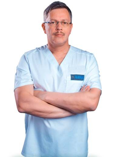
Honored Doctor of Ukraine, Head of the Plastic Surgery Center
Contact me
Plastic surgeon
Ukraine, Kyiv, Shchekavytska St., 9a
(Clinic "Nove Tilo")